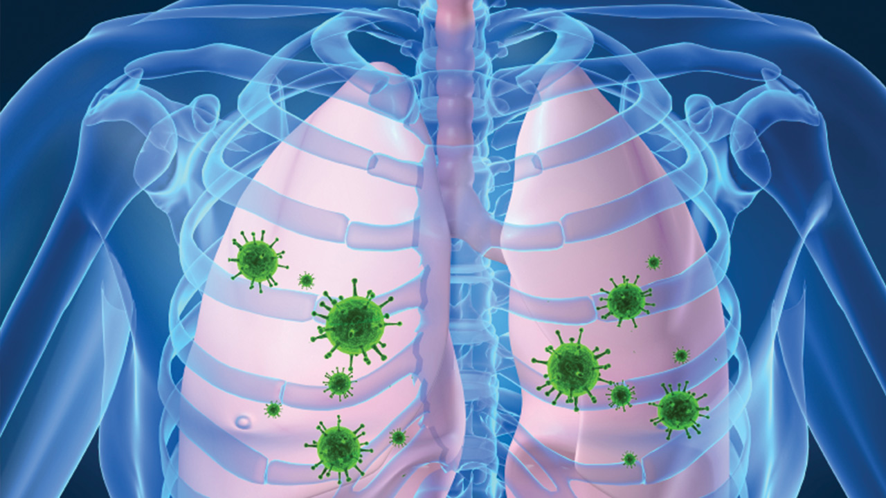
A ventilator is a machine that helps people breathe (ventilate). These machines are often used in hospitals as life support for patients who have difficulty breathing or who have lost all ability to breathe on their own. Mechanical ventilation may be either invasive or noninvasive (e.g. using a tight-fitting external mask). Invasive modes require the insertion of internal tubes/devices through endotracheal intubation or tracheostomy.
When Are Ventilators Used?
Mechanical ventilation is typically used on a temporary basis, such as during surgical procedures. When a patient is under general anesthesia, their normal breathing may be disrupted. A ventilator is used to ensure the patient continues to breathe while asleep during surgery.
A person with a serious lung disease or other medical condition that interferes with normal breathing may need to use a ventilator until they recover. This type of care is commonly provided in the intensive care unit (ICU) or critical care unit (CCU) of a hospital. Even if a patient with impaired lung function can breathe on their own, they may feel short of breath. Severe shortness of breath (dyspnea) can be very distressing and uncomfortable, but a ventilator can help ease the breathing process, allowing the patient to rest and heal.
Many diseases and other factors can affect lung function and cause difficulty breathing to the point that a person may need a ventilator to stabilize their condition. Examples include:
- Respiratory infections like pneumonia, influenza (flu) and coronavirus (COVID-19)
- Lung diseases like asthma, COPD (chronic obstructive pulmonary disease), cystic fibrosis and lung cancer
- Acute respiratory distress syndrome (ARDS)
- Damage to the nerves and/or muscles involved in breathing (can be caused by upper spinal cord injuries, polio, amyotrophic lateral sclerosis, myasthenia gravis, etc.)
- Brain injury
- Stroke
- Drug overdose
Patients who can’t breathe on their own at all also use ventilators while undergoing treatment for the underlying condition(s) that caused respiratory failure or respiratory arrest. Long-term ventilator care may be needed if a patient cannot regain the ability to breathe independently.
How Does a Ventilator Help a Person Breathe?
Invasive ventilators gently force normal air (or a mixture of air and added oxygen) through a breathing tube, into a patient’s airways and down into their lungs. Mechanical ventilation not only ensures that a patient receives sufficient oxygen but also helps move carbon dioxide, a waste gas, out of the lungs. A person who cannot breathe efficiently on their own may retain carbon dioxide in the body, which can accumulate and reach toxic levels.
It’s important to understand that ventilators are only used as life support; they do not treat or cure any medical conditions.
Intubation and Tracheostomy
Breathing tubes can be inserted using two different procedures: intubation or tracheostomy. During intubation, one end of the breathing tube is introduced through a patient’s nose or mouth and moved down into the throat until it enters the trachea. Breathing tubes placed in this manner are called endotracheal tubes. Tape or a specialized strap called an endotracheal tube holder is used to keep the breathing tube in place. Endotracheal tubes are typically used when a patient is on a ventilator for a short period of time.
When a patient requires mechanical ventilation for longer periods, the breathing tube may be placed in the windpipe using a procedure called a tracheostomy. During a tracheostomy, a surgeon makes an incision in the patient’s neck and trachea to create a hole called a stoma. The breathing tube is then inserted directly into the trachea via the stoma and secured using a strap that goes around the neck. A breathing tube placed like this is often referred to as a tracheostomy tube or “trach.”
Except in emergency situations where mechanical ventilation is needed immediately, both intubation and tracheostomy procedures are done in an operating room while the patient is under general anesthesia.
What Is Being on a Ventilator Like?
Mechanical ventilation isn’t usually painful, but the breathing tube may cause discomfort and take some getting used to. Patients often receive sedative and/or analgesic medications while on an invasive ventilator to minimize pain, agitation and anxiety. For patients who are awake, trach tubes tend to be more comfortable than endotracheal tubes. Being on a ventilator may also affect a person’s ability to talk. In some circumstances, a patient with a tracheostomy may still be able to speak.
Eating is impacted by invasive ventilation as well. Instead of taking food by mouth, a patient may require intravenous feeding (also known as IV feeding or parenteral nutrition) while on a ventilator. Tube feeding is common for those who are on a ventilator long term. With nasogastric (NG) feeding, the feeding tube is inserted through the nose, down the esophagus and down into stomach, while orogastric feeding involves placement of the tube through the mouth. Feeding tubes may also be placed through a surgically created hole (stoma) in the abdomen to introduce liquid nutrition directly into the stomach or small intestine.
For patients who are awake, being on a ventilator can significantly restrict mobility and activities. Those who require long-term ventilator care may be able to use a portable machine that allows for greater independence and freedom of movement.
A respiratory therapist usually tailors the ventilator’s settings to meet a patient’s unique needs and improve their comfort levels. For example, the machine can be set to “breathe” a certain number of times per minute. A patient may also be able to trigger the machine to initiate a breath. If a breath is not triggered in a certain timeframe, the machine will automatically act to ensure breathing continues.
In terms of ongoing care, breathing tubes must be suctioned out regularly to remove mucus that accumulates in the lungs. A nurse or respiratory therapist can perform this procedure, which typically triggers a brief bout of coughing and breathlessness.
Being on a Ventilator Comes with Health Risks
Patients who are on invasive ventilators require careful monitoring by medical professionals, including doctors, nurses and respiratory therapists. Chest X-rays and blood tests may be needed on a regular basis to ensure a patient is getting sufficient oxygen and not retaining excess carbon dioxide. Imaging and laboratory results will guide the patient’s care team in adjusting ventilator settings and other treatments as needed.
Attentive care will also help to detect and prevent complications of being on a ventilator. Typically, patients who need help breathing are already very ill and additional medical issues can jeopardize their recovery.
Ventilator-Associated Pneumonia
Ventilator-associated pneumonia (VAP) is a serious yet common complication of invasive ventilation. Bacteria can easily enter the body and lungs through the breathing tube, which also interferes with a patient’s ability to cough. Coughing is a natural mechanism that allows us to clear bacteria and other secretions from our lungs and airways.
Pneumonia can complicate a patient’s treatment, delay their recovery and even result in death. VAP is treated with antibiotics, but if the infection is caused by a drug-resistant type of bacteria, then much stronger antimicrobial drugs are needed to fight the infection.
Sinus Infection
Sinus infection is another risk of mechanical ventilation. This is more common in people who have an endotracheal tube. Sinus infections are also treated with antibiotics.
Ventilator-Associated Lung Injury
Various aspects of invasive ventilation may result in damage to tissues in and around the lungs. Therefore, it is important for medical professionals to carefully monitor patients who are on ventilators and adjust settings accordingly. Mechanical ventilation can cause injuries like pneumothorax (also known as a collapsed lung), a condition in which air leaks out of the lungs and into the pleural space between the lungs and the chest wall. Symptoms of pneumothorax include chest pain, low oxygen levels and shortness of breath.
Blood Clots
Using a ventilator also increases the risk of thrombotic events, such as deep vein thrombosis (DVT), pulmonary embolism (PE) and skin breakdown. These complications are more common in patients who have certain preexisting medical conditions and/or who remain in one position for extended periods, such as in bed or in a wheelchair.
Weaning From a Ventilator
Ventilators are lifesaving tools often used as a last resort. Because there are serious risks associated with mechanical ventilation, medical professionals usually try to discontinue ventilator use as soon as safely possible. The protocol for this is called weaning. The process involves spontaneous breathing trials during which a patient attempts to breathe with reduced or no support from the ventilator. Breathing trials are conducted under close supervision of one’s care team. Depending on the severity of a patient’s condition, weaning may take more than one try. Once a patient can breathe on their own, mechanical ventilation will be stopped. However, some patients never regain the ability to breathe independently and require long-term ventilation care.
